Dr Anne Maina treats a range of injuries affecting the lower and upper limbs.
Hover and Click on the trauma areas below for more information.
error: Content is protected !!
Clavicle
The clavicle, also known as the collarbone, is one of the bones of the shoulder joint that helps to provide strength and stability to the upper body.
A broken clavicle may occur after a fall onto an outstretched hand. It is one of the most common sports injuries, and may cause pain, an inability to raise the affected arm, and a grinding sensation when trying to move the arm. In some cases, a bump in the skin may appear at the fracture site.
In children, clavicle fractures are generally managed without surgery and heal well on their own after a brief period of immobilization. In adults, however, the risk of not healing completely is high, so surgery, followed by rehabilitation, is often offered to permit early range of movement and a faster return to work and sport.
Forearm
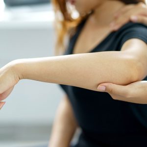
The forearm is made of two bones: the radius and the ulna. These two bones communicate with each other as well as the humerus proximally (toward the head) and carpal bones in the wrist distally (toward the fingers).
In children, these are two of the commonest bones sustain fractures. Most fractures occur toward the distal end of the bone, and occurs when the child – or adult – reaches out to break a fall.
While there are a variety of fracture patterns and dislocations in the forearem, children may occasionally additionally sustain incomplete (greenstick, buckle) fractures.
Humerus
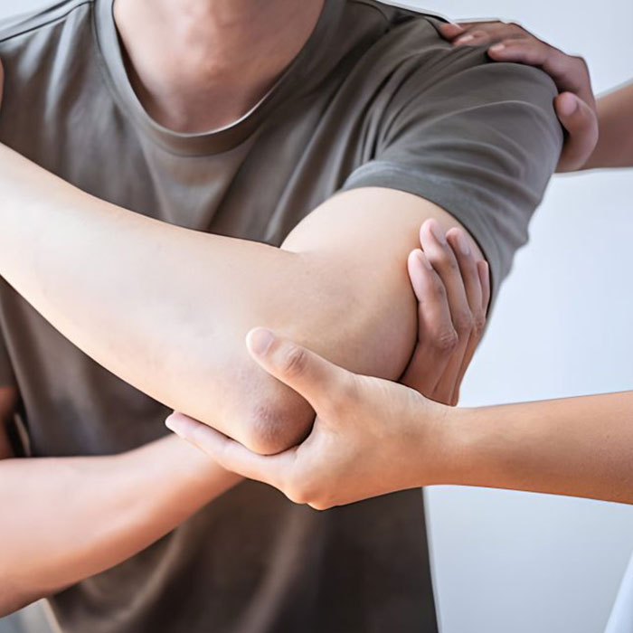
Fractures of the humerus (the long bone in the arm), are a common injury and can occur as the result of a fall or hard knock. In some cases, fractures in the humerus may heal without surgery. If, however, the fracture is particularly unstable, surgery may be recommended in order to realign and properly position the bone.
Surgery may involve the insertion of a nail, plates or an external fixator in the instance of severe or open injury. Physical rehabilitation is often an important part of the healing process following surgery.
Elbow
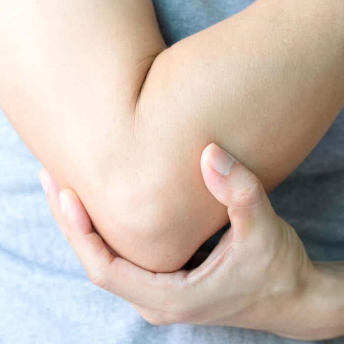
The elbow is a complex hinge joint formed by three bones: the humerus, radius and ulna. it is essential to upper limb function, but is prone to various types of acute and chronic injuries. These range from fractures, dislocations, torn ligaments to nerve entrapment syndromes, nerve injuries and so on. While some injuries heal well with conservative (non operative) measures and rehabilitation, others require surgery (emergent or otherwise) to permit an uncomplicated and functional recovery.
Surgery for more complex injuries aims at providing a stable joint with a preserved functional range of movement. It would often be preceded by advanced radiographic investigations (like a CT) to aid in surgical planning. Good outcomes are arrived at through a multidisciplinary team – with early range of movement exercises (supervised by the Occupational Therapist and or Physiotherapist) and a non-aggressive rehabilitation program.
Wrist / Distal Radius
The wrist is a complex region of the forearm, vulnerable to injury as a result of direct and indirect trauma. One of the most common wrist conditions is a fracture and/or dislocation. Fractures of the distal radius occur in children who suffer from falls on an outstretched hand, often when involved in recreation. On the other side of the spectrum, distal radius fractures often occur in middle-aged and elderly women suffering from osteoporosis.
These fractures are occasionally managed conservatively in a cast for several weeks. In fractures that appear significantly displaced, the broken bone is realigned in the operating room, and a cast is appplied, with or without wires. In older individuals, fractures are reduced and a plate and screws is inserted.
Occasionally there is associated damage to the ligaments of the wrist. In those instances, dislocations of the small bones in the wrist require stabilization of the small bones of the wrist.
Hip
Joint replacement surgery is a procedure in which the doctor treats severe arthritis by surgically replacing it with an artificial joint typically made from metal and plastic components.
This surgery is usually done when all other treatment options for joint pain have failed to provide adequate pain relief.
Dr. Maina is one of a handful of surgeons registered and trained to use the (Anterior Minimally Invasive Surgery) technique for hip replacements in South Africa.
The AMIS approach is unique as it creates less surgical trauma than other surgical techniques for hip replacements.
Femur
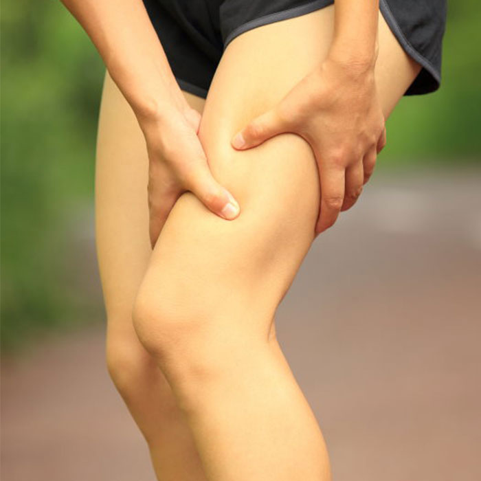
The femur is the strongest bone in the body but is often fractured in high energy accidents, but in older osteopaenic or osteoporotic people, are broken by low energy fractures. The symptoms are pain and inability to bear weight on the leg. Femur fractures generally require surgery to stabilise the leg, restore mobility, function and allow rehabilitation.
In the elderly, occasionally, the fracture involves the neck of the femur. In this case, there may be a need for total or partial hip replacement surgery. When the fracture involves the trochanteric area, a femur nail is inserted to stabilise the femur and allow rehabilitation and mobilization.
Fractures involving the shaft (middle) or lower part of the femur, are often stabilised using a nail and/or a plate and scews.Surgery is often performed within a day or two of the injury to decrease the length of stay in hospital, the risk of complications such as blood clots, lung complications and allow early walking. Sometimes fractures may occur around hip or knee prosthesis (hip replacement or knee replacement implants).
Patella
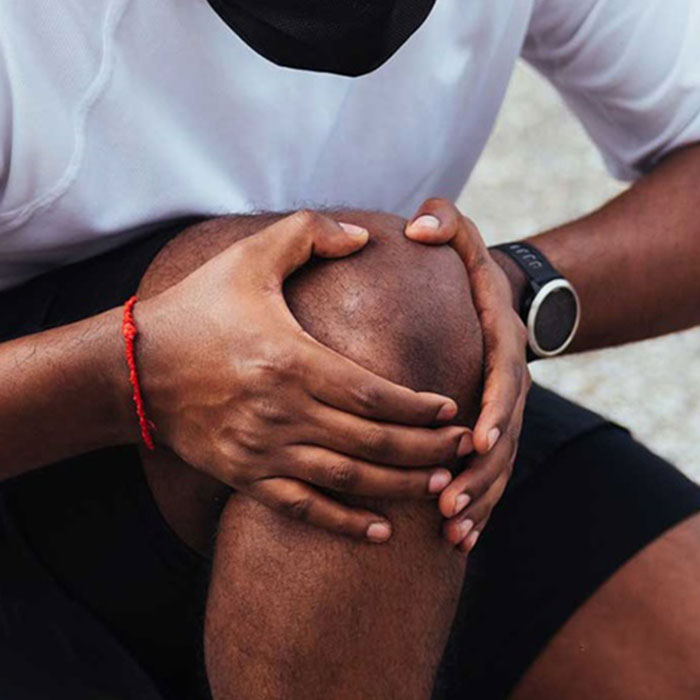
Knee dislocations are major injuries that can occur with high energy injuries. They often involve disruptions of the ligaments and cartilage (meniscus) of the knee. Aside from fractures, often, dislocations involve damage to blood vessels and/or nerves of the leg.
Ligaments damaged by a dislocation may include the anterior cruciate ligament (ACL), posterior cruciate ligament (PCL) and collateral ligaments. Integrity of these structures is often determined using an MRI scan. The MRI, although an expensive investigation, is invaluable in providing information as to the extent of ligament, cartilage, bone and soft tissue damage in the knee.
Reconstruction (or repair) of the ligaments is often done two or more weeks after the injury to allow joint capsule tears to heal and allow the patient to gain some range of movement in the affected knee. Reconstruction, or repair, is conducted arthroscopically – also referred to as ‘keyhole surgery’ – leaving minimal scarring. One’s own ligaments (hamstring, quadriceps etc) are usually used as grafts due to the low risk of infection, low risk of failure and the good rate of incorporation of the reconstructed ligament.
Whenever a knee dislocation occurs, there is a risk of excessive swelling in the muscles of the leg – a complication called ‘compartment syndrome’. Symptoms of this potentially limb and life threatening condition include: severe pain, pins and needles, paralysis and a pale extremity. These symptoms are often managed with decompression of the muscle compartments in the leg – an emergency procedure to open the damaged muscle compartments, called a fasciotomy.
Tibia
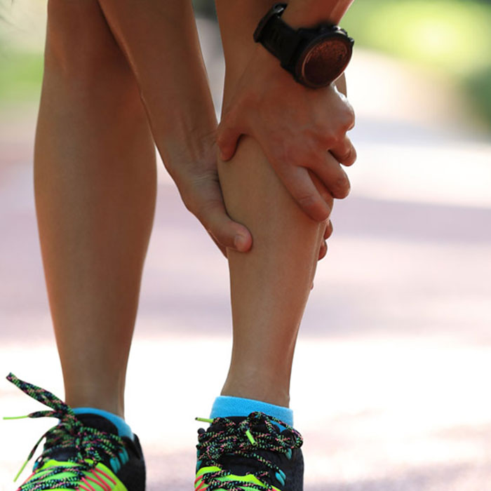
The tibia is one of the most commonly fractured bones in the body. Often a tibia fracture occurs at the same time, but not necessarily at the same level, as a fibula fracture. This may occur with both low and high energy trauma. The most common sites of a tibia fracture are the tibial shaft (middle) and tibia plateau (upper end). Symptoms of a tibial fracture include pain, inability to bear weight on the affected leg, instability and sometimes a visible deformity.
Treatment will include surgery that may require one or more of the following to be applied in the operating theatre: a cast, insertion of a plate, insertion of screws and/or insertion of a nail. In open fractures, the wound will also need to be cleaned (debridement).
As with knee dislocations, whenever a tibia fracture occurs, there is a risk of excessive swelling in the muscles of the leg – a complication called ‘compartment syndrome’. Symptoms of this potentially limb and life threatening condition include: severe pain, pins and needles, paralysis and a pale extremity. These symptoms are often managed with decompression of the muscle compartments in the leg – an emergency procedure to open the damaged muscle compartments, called a fasciotomy.
Ankle
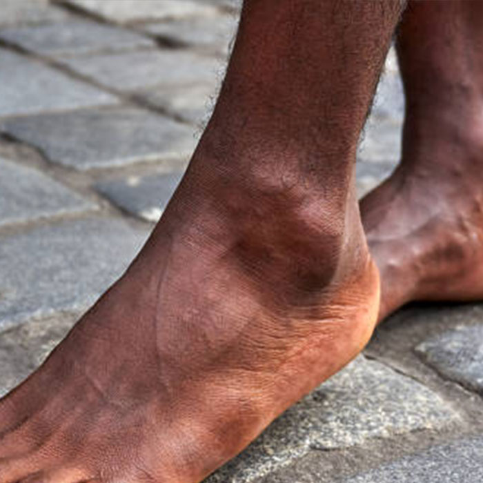
The most common ankle injury include ankle sprains, fractures and dislocations. An x-ray (and sometimes a CT scan and/or MRI) will be done to confirm the diagnosis and extent of injury. Ankle fractures are often closed injuries, but open injuries and unstable ankle fracures often require surgery.
Often, when someone presents after more than a few hours to the doctor, the ankle can be very swollen. Should the injury require surgery, one may be required to be admitted to the ward to elevate the ankle and use ice packs to allow the swelling to settle prior to surgery. This is to reduce the risk of skin complications often reported with swollen soft tissues at the time of surgery.
After surgery to stabilise a fractured ankle, you will be required to walk with crutches and wear a cast or moon boot to provide further support and stability to the joint.